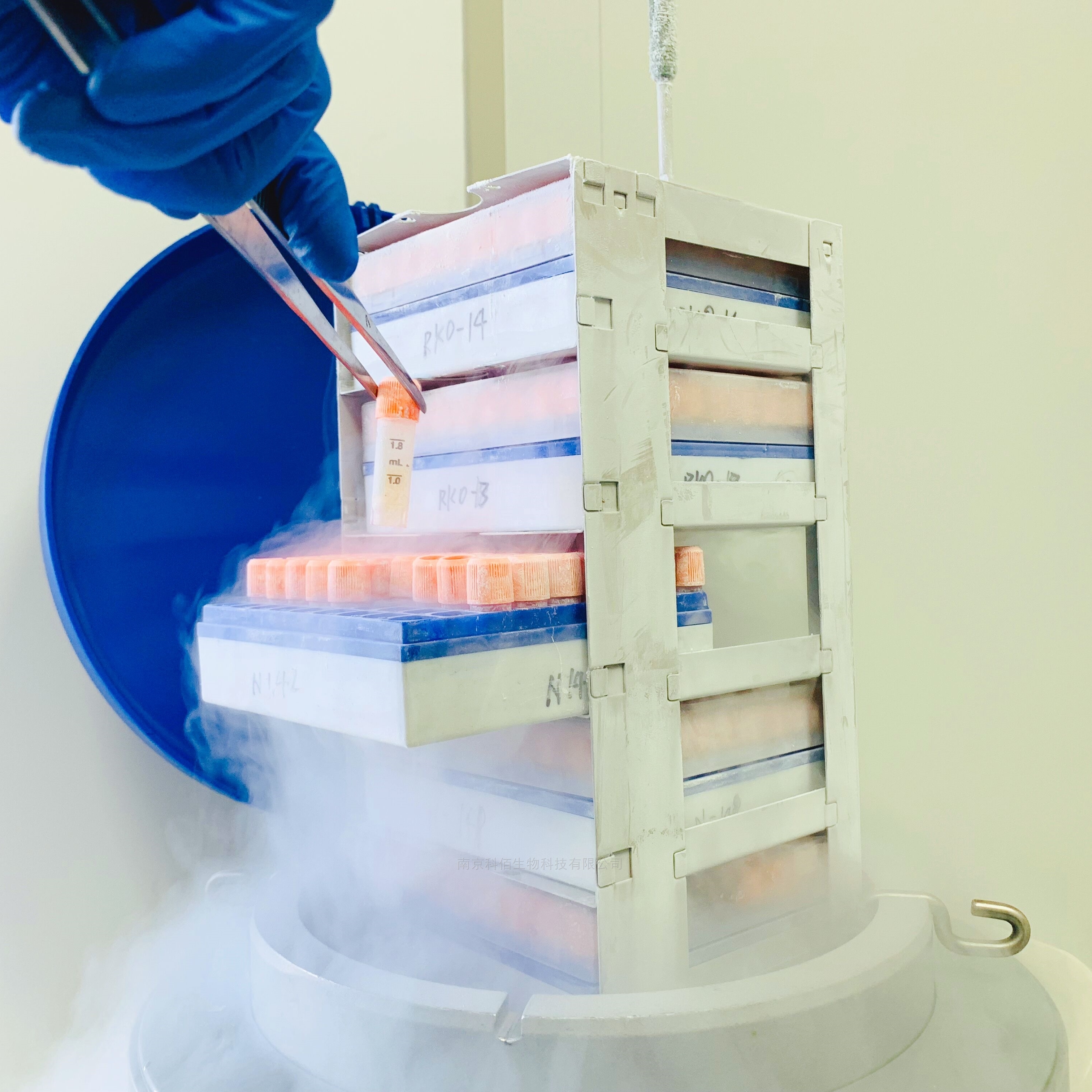
| 名称 | PD1/NFAT-Luc/Jurkat |
| 型号 | CBP74018 |
| 报价 |  |
| 特点 | PD1/NFAT-Luc/Jurkat,冻存条件:90% FBS+10% DMSO; |
产品搜索
相关文章
联系我们
电话:4008750250
号码:
手机:18066071954
地址:南京市栖霞区纬地路9号
Email: zhangxiangwen@cobioer.com
产品展示 / PRODUCTS

- 详细内容
| I. Background | |
| 肿瘤细胞可以借助免疫检查点受体逃避机体免疫系统的识别和杀伤,因此阻断免疫检 查点受体可能是一种广泛有效的治疗方法。目前,抗 PD-1/PD-L1 抗体虽然比较 成熟,与抗 CTLA4 抗体类似,但由于存在耐药性,患者的总体有效率较低,因此寻找新 的治疗靶点迫在眉睫。 Programmed Cell Death Protein 1 (PD-1),一种在激活的 T 细胞上表达的受体,与其配 体 PD-L1 和 PD-L2 结合,负向调节免疫反应。 PD-1 配体存在于大多数癌症中,PD-1:PD- L1/2 相互作用会抑制 T 细胞活性,并使癌细胞逃避免疫监视。 PD1/PDL1 信号转导通路是 肿瘤免疫抑制的重要组成部分,可以抑制 T 淋巴细胞的兴奋,增强肿瘤细胞的免疫耐受, 从而实现肿瘤免疫逃逸。PD1 与 PDL1 结合可以减弱 T 细胞介导的免疫监视,导致免疫反 应缺失,甚至导致 T 细胞凋亡。PD1/PDL1 抑制剂可解除抗肿瘤 T 细胞的免疫抑制,从而 导致 T 细胞增殖并渗透到肿瘤微环境中并诱导抗肿瘤反应。PD-1:PD-L1/2 通路还参与调节 自身免疫反应,使这些蛋白质成为多种癌症以及多发性硬化症、关节炎、狼疮和 I 型糖尿 病的有希望的治疗靶点。磷酸酶 SHP2 是 T 细胞功能的关键调节因子,它介导 TCR 下游的激活信号以及 PD-1 下游的抑制信号。 | |
| II. Description | |
PD1 NFAT-Luc Jurkat 报告基因药靶模型很好的模拟了体内 PD1&PDL1/PDL2 的信号转 导过程,原理见下图所示。  Figure 1. PD1 NFAT-Luc Jurkat 细胞模型原理图 | |
| III. Introduction | |
| Host Cell: | Jurkat |
| Expressed gene: | PD1、NFAT-Luciferase |
| Stability: | 32 passages (in-house test, that not means the cell line will be instable beyond the passages we tested.) |
| Synonym(s): | Programmed cell death 1, PDCD1, PD-1, PD1, SLEB2, CD279, HPD-L, PD1/NFAT, PD-1 NFAT, PD-1/NFAT |
| Freeze Medium: | 90% FBS+10% DMSO |
| Culture Medium: | RPMI-1640+10%FBS+800μg/ml Hygromycin+1μg/ml Puromycin |
| Mycoplasma Testing: | Negative |
| Storage: | Liquid nitrogen |
| Application(s): | Functional(Report Gene) Assay |
| IV. Description of Host Cell Line | |
| Organism: | Homo sapiens, human |
| Tissue: | Peripheral blood |
| Disease: | Acute T cell leukemia |
| Morphology: | Lymphoblast |
| Growth Properties: | Suspension |
| Ⅴ. Representative Data | |
| |
Figure 2. Recombinant PD1/NFAT-Luc/Jurkat stably expressing PD1. | |
Figure 3. Detect Luciferase assay by Ultra Luciferase Detection Kit CBPH0001(we strongly suggest to purchase from Cobioer).Dose response of Anti-PD-1 Neutralizing Antibody in PD1 NFAT-Luc Jurkat(C16) with PDL1 TCR Activator CHO (C5). | |
| |
Figure 4. Detect Luciferase assay by Ultra Luciferase Detection Kit CBPH0001(we strongly suggest to purchase from Cobioer). Dose response of Anti-PD-1 Neutralizing Antibody in PD1 NFAT-Luc Jurkat(C16) with PDL1 TCR Activator CHO (C5). | |
Figure 5. Detect Luciferase assay by Ultra Luciferase Detection Kit CBPH0001(we strongly suggest to purchase from Cobioer). Dose response of Anti-PD1 Neutralizing Antibody in PD1 NFAT-Luc Jurkat(C16) with PDL2 TCR Activator CHO(C1).. | |
Figure 6. Detect Luciferase assay by Ultra Luciferase Detection Kit CBPH0001(we strongly suggest to purchase from Cobioer). Dose response of PD1 Agonist Antibody in PD1 NFAT-Luc Jurkat(C16) with FcγR aAPC Cell(C1). | |


